
Plain FilmsOf the AbdomenAn Approach
Plain FilmsOf the AbdomenAn Approach
© William Herring, MD, FACR

Lecture Map
Normal Gas Pattern
Obstruction Series
Abnormal Gas Pattern
Aunt Minnies
Extraluminal Air
What to look for
Soft Tissue Masses
Calcifications

What to Examine
Gas pattern
Extraluminal air
Soft tissue masses
Calcifications


Normal Gas Pattern
Stomach
Always
Small Bowel
Two or three loops of non-distended bowel
Normal diameter = 2.5 cm = 1 US quarter
Large Bowel
In rectum or sigmoid – almost always


Gas instomach
Gas in a fewloops ofsmall bowel
Gas inrectum orsigmoid
Normal Gas Pattern

Normal Fluid Levels
Stomach
Always (except supine film)
Small Bowel
Two or three levels possible
Large Bowel
None normally


Erect Abdomen
Alwaysair/fluid levelin stomach
A fewair/fluidlevels insmall bowel

Large vs. Small Bowel
Large Bowel
Peripheral
Haustral markings don't extendfrom wall to wall
Small Bowel
Central
Valvulae extend across lumen
Maximum diameter of 2"


Complete AbdomenObstruction Series
Supine
Prone or lateral rectum
Erect or left decubitus
Chest - erect or supine

Complete AbdomenSupine
Looking for
Scout film for gaspattern
Calcifications
Soft tissue masses
Substitute – none


Complete AbdomenProne
Looking for
Gas in rectum/sigmoid
Gas in ascending anddescending colon
Substitute – lateralrectum


Complete AbdomenErect
Looking for
Free air
Air-fluid levels
Substitute – leftlateral decubitus


Left Lateral Decubitus View


Amateur Style

Complete AbdomenErect Chest
Looking for
Free air
Pneumonia at bases
Pleural effusions
Substitute – supinechest


Abnormal Gas Patterns
Functional Ileus
Localized (Sentinel Loops)
Generalized adynamic ileus
Mechanical Obstruction
SBO
LBO

Laws of the Gut
Loops proximal to an obstruction willbe dilated
Loops distal to an obstruction will bedecompressed or airless
Most dilated loop will either be:
Most distended to start
Bowel just proximal to the obstruction

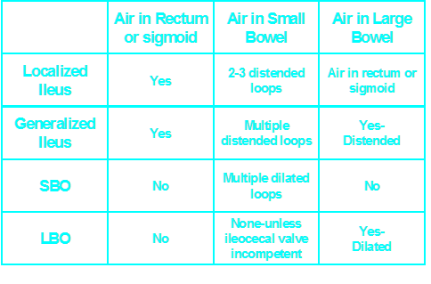

One or two persistently dilated loops oflarge or small bowel
Gas in rectum or sigmoid
Localized IleusKey Features

Sentinel Loops

Supine

Prone


PancreatitisUlcer
Diverticulitis
Cholecystitis
Appendicitis
UlcerUreteral calculus
Sentinel Loops

Localized IleusPitfalls
May resemble earlymechanical SBO
Clinical course
Get follow-up


Gas in dilated small bowel and largebowel to rectum
Long air-fluid levels
Only post-op patients havegeneralized ileus!
Generalized IleusKey Features


Generalized Adynamic Ileus
Supine
Erect

Is It An Ileus?
Is the patient immediately post-op?
Are the bowel sounds absent orhypoactive?
If “no,” then it isn’t an ileus
Patients don’t present to the ER with ageneralized adynamic ileus!

Mechanical SBOKey Features
Dilated small bowel
Fighting loops
Little gas in colon, especially rectum
Key: disproportionate dilatation of SB


SBO


Mechanical SBOCauses
Adhesions
Hernia*
Volvulus
Gallstone ileus*
Intussusception
*Cause may be visible on plain film

Mechanical SBOPitfalls
Early SBO mayresemblelocalized ileus -get F/O


Mechanical LBOKey Features
Dilated colon to point of obstruction
Little or no air in rectum/sigmoid
Little or no gas in small bowel, if…
Ileocecal valve remains competent

LBO

Supine

Prone

Mechanical LBOCauses
Tumor
Volvulus
Hernia
Diverticulitis
Intussusception

Mechanical LBOPitfalls
Incompetent ileocecal valve
Large bowel decompresses into smallbowel
May look like SBO
Get BE or follow-up


Carcinoma of Sigmoid – LBO –Decompressed into SB

Prone

Supine


Aunt Minnies


Air inbiliarytree
SBO
Gallstone
Gallstone Ileus


Post-op C-sectionAdynamic Ileus
Soft tissuemass inpelvis
Midlinesutures
Dilatedsmall andlarge boweldisplacedfrom pelvis


Sigmoid Volvulus


Mesenteric Occlusion

Abnormal Gas PatternsIleus and Obstruction
Localized ileus
Generalized ileus
Mechanical SBO
Mechanical LBO

Extraluminal AirFree Intraperitoneal Air

Signs of Free Air
Air beneath diaphragm
Both sides of bowel wall
Falciform ligament sign


ERECT
Crescentsign
Free Intraperitoneal Air


Free Intraperitoneal Air
Air on both sides ofbowel wall – Rigler’sSign


Free Intraperitoneal Air
FalciformLigamentSign

Football sign

Free AirCauses
Rupture of a hollow viscus
Perforated ulcer
Perforated diverticulitis
Perforated carcinoma
Trauma or instrumentation
Post-op 5–7 days
NOT perforated appendix


Air in Lesser Sac

Extraperitoneal Air


Intermission
There is no such thing as a “non-specific”gas pattern
It is either normal or abnormal
Adynamic Ileus is overcalled
Only occurs in post-op patients
Take notes in conference

Soft Tissue Masses

Soft Tissue Masses
Hepatosplenomegaly
Plain films poor for judging liver size
Tumor or cyst
Bowel displacement
Paucity of gas
Pad sign
•Extrinsic compression of bowel


Splenomegaly


Myomatous Uterus


Bladder Outlet Obstruction – pre- and post- cath
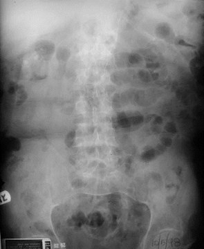
Hourslater

Hourslater


Mass in Cologastric Space - Pancreatic Pseudocyst


Right Renal Cyst


RLQ Abscess


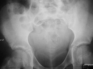
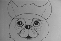
Free Peritoneal Fluid- Bladder Ears

AbdominalCalcifications
AbdominalCalcifications

Abdominal CalcificationsPatterns
Rimlike
Linear or track-like
Lamellar
Cloudlike

Rimlike Calcification
Wall of a hollow viscus
Cysts
Renal cyst
Aneurysms
Aortic aneurysm
Saccular organs e.g. GB
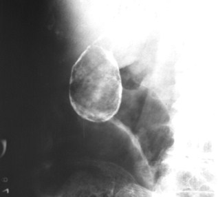
Porcelain Gallbladder

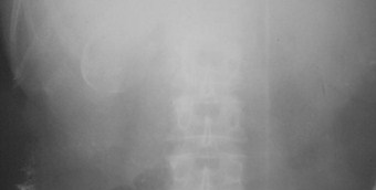
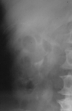
Renal Cyst
Gallbladder Wall

Linear or Track-like
Walls of a tube
Ureters
Arterial walls

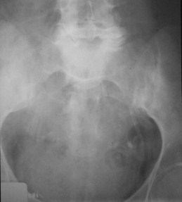
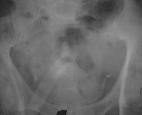
Atherosclerosis
Calcification Vas Deferens

Lamellar or Laminar
Formed in lumen of a hollow viscus
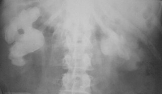
Renal stones
Gallstones
Bladder stones

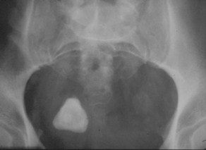
Stone in Ureterocoele
Staghorn Calculi

Cloudlike, Amorphous, Popcorn
Formed in a solid organ or tumor
Leiomyomas of uterus
Ovarian cystadenomas

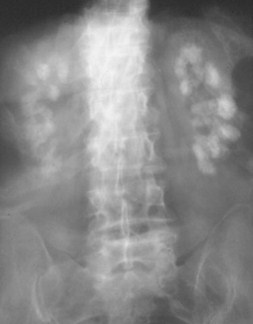
Nephrocalcinosis
Myomatous Uterus
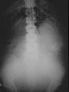

Unknowns

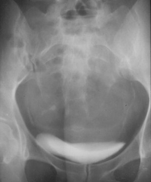
Dermoid

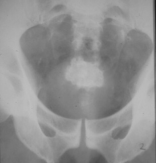
Jackstone Calculus


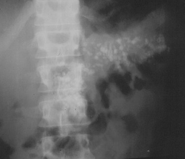
Calcific Pancreatitis

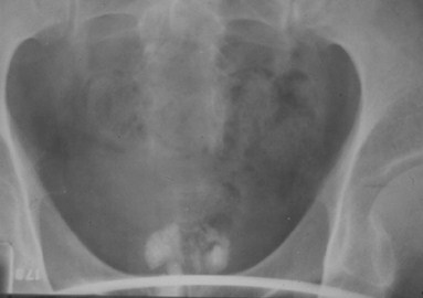
Prostatic Calcification

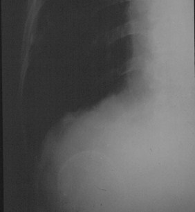
Renal cyst

Porcelain Gallbladder

What to Examine
Gas pattern
Extraluminal air
Soft tissue masses
Calcifications


